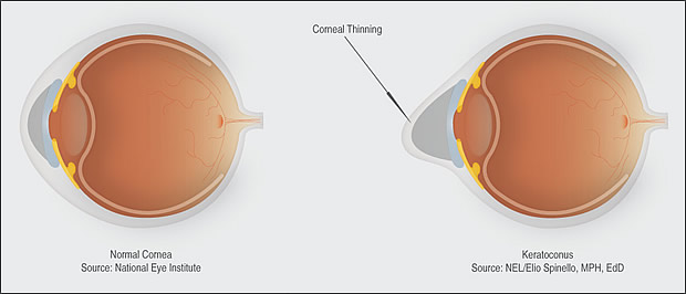
Keratoconus is an eye disorder characterized by an irregular corneal surface (cone-shaped) resulting in blurred and distorted images. Keratoconus literally means cone-shaped cornea (American Academy of Ophthalmology, 2011).

The cornea is the window and outer surface of the eye. When you are interpreting an image, light travels through the cornea past the lens to the retina and then the brain to form a visual image. The normal corneal surface is smooth and aspheric and flattens towards the edges. Light rays passing through it moves in an undistorted manner to the retina to project a clear image to the brain. This is the typical normal working cornea.
Keratoconus is a very slow progressive eye condition that affects the cornea. The normally round, dome-shaped cornea weakens and thins, causing a cone-like bulge to develop. The regular curvature of the cornea becomes irregular, resulting in increasing nearsightedness (myopia) and astigmatism that have to be corrected with special glasses or contact lenses. Since the cornea is responsible for refracting most of the light coming into the eye, corneal abnormalities can result in significant visual impairment, making simple tasks like driving or reading books.
Symptoms of Keratoconus (EYE FACTS AAO worksheet, 2010)
- mild burning
- glare at night
- irritable eye
- sensitivity to light
- some distortion of vision
According to the AAO, about 1/ 2000 people will develop keratoconus. Most people will have a mild or moderate form of the disease. Less than 10% of people with keratoconus will develop the most severe form. It typically is diagnosed in the late teens or twenties. However, many people have been diagnosed in their mid to late thirties. It is common for one eye to precede faster than the other and the eyes may go for long periods of time without any change and then change dramatically over a period of months.


